xylose chair conformation PDB 1OD8 was discovered to feature the acidbase catalytic residue Glu128 in the deprotonated state whereas the nucleophile Glu 236 was found in protonated. The detection of xylose cyclic phosphonate as the turnover product reveals significant new details about the AXS-catalyzed reaction and supports the proposed retroaldol-aldol mechanism of catalysis.
Xylose Chair Conformation, And by Mohr 13 namely the chair and boat forms could be extended to pyranoid sugars. Solved Expert Answer to Draw the following xylose derivative in a chair conformation and then consider the conformation of the ester substituents. Draw the product s from Killiani-Fischer reaction of D-ribose and L-Xylose.
 Synthesis Of 2 3 O Benzyl Ribose And Xylose And Their Equilibration Sciencedirect From sciencedirect.com
Another Article :
What is the full name of the monosaccharide shown below. Hydrogenation of the C4 keto group by NADH assisted by Tyr147 as catalytic proton donor yields UDP-xylose adopting the relaxed 4C1 chair conformation step 3. The resulting UDP-4-keto-pentose adopts a flattened 4 C 1 chair Fig. Organic Chemistry 3rd Edition. A cyclohexane molecule in chair conformation.
For each chair conformer add the energy of all the groups on axial position.
Do you mean D-xylose. Besides its applications in bioenergy and biosynthesis β-D-xylose is a very simple monosaccharide that exhibits relatively high rigidity. Structure of the XylFII-LytSN-xyl complex. The detection of xylose cyclic phosphonate as the turnover product reveals significant new details about the AXS-catalyzed reaction and supports the proposed retroaldol-aldol mechanism of catalysis. Draw the chair conformation of the following.
 Source: fi.m.wikipedia.org
Tiedosto D Xylose Svg Wikipedia The reactive 4-keto group is placed suitably for stereospecific hydride transfer from NADH under general acid catalytic assistance from Tyr 147. And now the stabilities. The data suggest that this observation is unlikely to be due to an unfavorable equilibrium but rather results from substrate inhibition by the most stable chair conformation of UDP-D-xylose. Structure of the XylFII-LytSN-xyl complex. 2- what are the chair conformation for D-xylulose. The C2-C3-C5-O square forms a plane whereas the C1 and C4 atoms take up a higher or lower position thus yielding the two possible chairs 1C 4 and 4C 1.
 Source: cell.com
L Arabinose Binding Isomerization And Epimerization By D Xylose Isomerase X Ray Neutron Crystallographic And Molecular Simulation Study Structure The occurrence in 23-di-O-methyl- d -xylose of a lower frequency band near 3530 cm 1 suggests the presence of hydrogen bonds between cis meta diaxial groups which would be possible only if the 1C. PDB 1OD8 was discovered to feature the acidbase catalytic residue Glu128 in the deprotonated state whereas the nucleophile Glu 236 was found in protonated. Like the cyclohexane ring adopt a low-energy conformation that looks like a chair. 1- What is the alpha and beta haworth conformation of D-xylulose. In the first conformer we have two chlorines in axial positions so the total steric strain is. They found that the chair conformations of the D-glucopyranose units satisfactorily explained the spacings in the X-ray diagram of cellulose whereas the boat con formations did not.
 Source: masterorganicchemistry.com
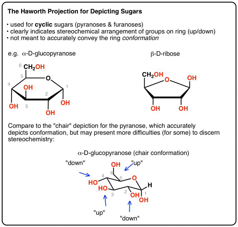
The Haworth Projection Master Organic Chemistry Fischer Projections Haworth Structures and Chair Conformers The acyclic structure of a sugar is commonly drawn as a Fischer projection. For α-glucose β-glucose and β-mannose 729 rotamers were generated for each pucker resulting in 27 702 conformations for each of these six-carbon monosaccharides. They found that the chair conformations of the D-glucopyranose units satisfactorily explained the spacings in the X-ray diagram of cellulose whereas the boat con formations did not. The reactive 4-keto group is placed suitably for stereospecific hydride transfer from NADH under general acid catalytic assistance from Tyr 147. A trajectory initiated for the wild-type enzymesubstrate complex with the proximal xylose ring bound at the 1 subsite adjacent to the scissile glycosidic bond in the 4 C 1 chair conformation shows spontaneous transformation to the 25 B boat conformation and potential of mean force calculations indicate that the boat is 30 kJ mol 1 lower in free energy than the chair. The resulting UDP-4-keto-pentose adopts a flattened 4 C 1 chair Fig.
 Source: pubs.rsc.org
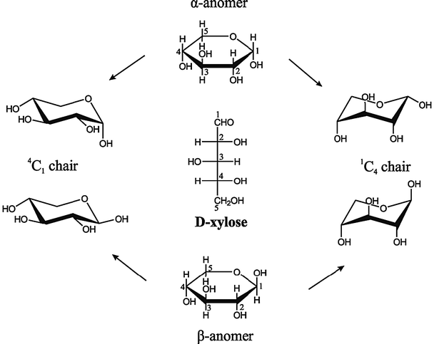
Conformations Of D Xylose The Pivotal Role Of The Intramolecular Hydrogen Bonding Physical Chemistry Chemical Physics Rsc Publishing Doi 10 1039 C3cp52345d Because many compounds feature structurally similar six-membered rings. Although D-glucose has a strong preference for the 4C 1. Like the cyclohexane ring adopt a low-energy conformation that looks like a chair. In the first conformer we have two chlorines in axial positions so the total steric strain is. Arg277 which is positioned by a salt-link interaction with Glu120 closes up the catalytic site and prevents release of the UDP-4-keto-pentose and NADH intermediates. As such it provides the best.
 Source: chegg.com
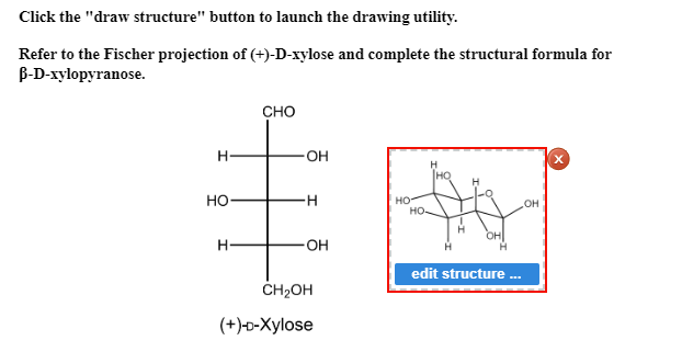
Solved Click The Draw Structure Button To Launch The Chegg Com By the NTD and CTD of XylFII the pyranose ring of the D-xylose molecule adopts a chair conformation and residues from both ter-minal domains of XylFII form hydrophobic and hydrogen-bonding Fig. The data suggest that this observation is unlikely to be due to an unfavorable equilibrium but rather results from substrate inhibition by the most stable chair conformation of UDP-D-xylose. A Structure of the WT XylFII-LytSN complex with bound D-xylose XylFII-LytSN-xyl. The occurrence in 23-di-O-methyl- d -xylose of a lower frequency band near 3530 cm 1 suggests the presence of hydrogen bonds between cis meta diaxial groups which would be possible only if the 1C. By the NTD and CTD of XylFII the pyranose ring of the D-xylose molecule adopts a chair conformation and residues from both ter-minal domains of XylFII form hydrophobic and hydrogen-bonding Fig. The reactive 4-keto group is placed suitably for stereospecific hydride transfer from NADH under general acid catalytic assistance from Tyr 147.

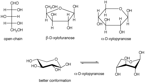
2 252 It is noteworthy that the complex with the corresponding lactam 121 Scheme 30. The reactive 4-keto group is placed suitably for stereospecific hydride transfer from NADH under general acid catalytic assistance from Tyr 147. A cyclohexane molecule in chair conformation. Draw the 2 chair conformation of the following pyranose forms of beta d mannose alpha D xylose beta D galactose alpha D fructose and identify which of the conformations has the lowest energy. Abstract and Figures. The UDP-xylose product is in a relaxed 4 C 1 conformation.
 Source: publishing.cdlib.org
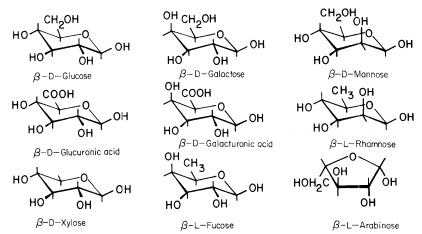
The Molecular Biology Of Plant Cells D0e311 B acetic anhydride in pyridine b D-galactose b D-allose. Solved Expert Answer to Draw the following xylose derivative in a chair conformation and then consider the conformation of the ester substituents. For each chair conformer add the energy of all the groups on axial position. The UDP-xylose product is in a relaxed 4 C 1 conformation. The resulting UDP-4-keto-pentose adopts a flattened 4 C 1 chair Fig. Structure of the XylFII-LytSN-xyl complex.

2 And now the stabilities. Draw the second chair conformation ring-flip-check this post if not sure. In organic chemistry cyclohexane conformations are any of several three-dimensional shapes adopted by molecules of cyclohexane. Other principal conformations of pyranoses are halfchair H boat B and skew S conformation which are named as indicated. The resulting UDP-4-keto-pentose adopts a flattened 4 C 1 chair Fig. Kersters-Hilderson H Claeyssens M van Doorslaer E de Bruyne CK.
 Source: researchgate.net
Scheme 1 Fisher Projection Of D Xylose Centre And Haworth Projections Download Scientific Diagram On the basis of A1 Get Best Price Guarantee. Like the cyclohexane ring adopt a low-energy conformation that looks like a chair. The occurrence in 23-di-O-methyl- d -xylose of a lower frequency band near 3530 cm 1 suggests the presence of hydrogen bonds between cis meta diaxial groups which would be possible only if the 1C. The reactive 4-keto group is placed suitably for stereospecific hydride transfer from NADH under general acid catalytic assistance from Tyr 147. The data suggest that this observation is unlikely to be due to an unfavorable equilibrium but rather results from substrate inhibition by the most stable chair conformation of UDP-D-xylose. Solved Expert Answer to Draw the following xylose derivative in a chair conformation and then consider the conformation of the ester substituents.
 Source: web.pdx.edu
Quiz 25 By the NTD and CTD of XylFII the pyranose ring of the D-xylose molecule adopts a chair conformation and residues from both ter-minal domains of XylFII form hydrophobic and hydrogen-bonding Fig. 252 It is noteworthy that the complex with the corresponding lactam 121 Scheme 30. These structures make it easy to show the configuration at each stereogenic center in the molecule without using wedges and dashes. On the basis of A1 Get Best Price Guarantee. Abstract and Figures. Because many compounds feature structurally similar six-membered rings.

Solved 1 What Is The Alpha And Beta Haworth Conformation Of D Xylulose 2 What Are The Chair Conformation For D Xylulose Course Hero Lowest energy conformations have the largest nonhydrogen subsstituents occupying the lowest energy equatorial position. Because many compounds feature structurally similar six-membered rings. The data suggest that this observation is unlikely to be due to an unfavorable equilibrium but rather results from substrate inhibition by the most stable chair conformation of UDP-D-xylose. The reactive 4-keto group is placed suitably for stereospecific hydride transfer from NADH under general acid catalytic assistance from Tyr 147. The resulting UDP-4-keto-pentose adopts a flattened 4 C 1 chair Fig. The resulting UDP-4-keto-pentose adopts a flattened 4 C 1 chair Fig.
 Source: researchgate.net
A Equatorial And Axial Directions Of The Ring Shown With E And A Download Scientific Diagram And now the stabilities. In the first conformer we have two chlorines in axial positions so the total steric strain is. Lowest energy conformations have the largest nonhydrogen subsstituents occupying the lowest energy equatorial position. The detection of xylose cyclic phosphonate as the turnover product reveals significant new details about the AXS-catalyzed reaction and supports the proposed retroaldol-aldol mechanism of catalysis. Draw the 2 chair conformation of the following pyranose forms of beta d mannose alpha D xylose beta D galactose alpha D fructose and identify which of the conformations has the lowest energy. For each chair conformer add the energy of all the groups on axial position.
 Source: cell.com
L Arabinose Binding Isomerization And Epimerization By D Xylose Isomerase X Ray Neutron Crystallographic And Molecular Simulation Study Structure These structures make it easy to show the configuration at each stereogenic center in the molecule without using wedges and dashes. The detection of xylose cyclic phosphonate as the turnover product reveals significant new details about the AXS-catalyzed reaction and supports the proposed retroaldol-aldol mechanism of catalysis. Both of these were found in the 4 C 1 chair conformation in their complexes with xylanase Xyn10A from Strepomyces lividans a retaining endoxylanase GH 10. A B-D - galactopyranose b B-D mannopyranose 7. The resulting UDP-4-keto-pentose adopts a flattened 4 C 1 chair Fig. Do you mean D-xylose.
 Source: researchgate.net
Open And Cyclic Form Of D Xylose With 4 C1 And 1 C4 Chair Download Scientific Diagram Because many compounds feature structurally similar six-membered rings. For most of the compounds studied the spectra are consistent with the predominance of the C1 conformation of the pyranose ring but there is evidence of some departures from the ideal chair shape. 252 It is noteworthy that the complex with the corresponding lactam 121 Scheme 30. Although D-glucose has a strong preference for the 4C 1. Arg277 which is positioned by a salt-link interaction with Glu120 closes up the catalytic site and prevents release of the UDP-4-keto-pentose and NADH intermediates. Because many compounds feature structurally similar six-membered rings.
 Source: masterorganicchemistry.com
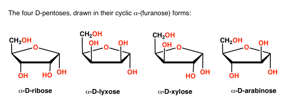
The Haworth Projection Master Organic Chemistry Although D-glucose has a strong preference for the 4C 1. For most of the compounds studied the spectra are consistent with the predominance of the C1 conformation of the pyranose ring but there is evidence of some departures from the ideal chair shape. In organic chemistry cyclohexane conformations are any of several three-dimensional shapes adopted by molecules of cyclohexane. Because many compounds feature structurally similar six-membered rings. Structure of the XylFII-LytSN-xyl complex. The resulting UDP-4-keto-pentose adopts a flattened 4 C 1 chair Fig.
 Source: researchgate.net
A Equatorial And Axial Directions Of The Ring Shown With E And A Download Scientific Diagram Draw the product s from Killiani-Fischer reaction of D-ribose and L-Xylose. Although D-glucose has a strong preference for the 4C 1. A trajectory initiated for the wild-type enzymesubstrate complex with the proximal xylose ring bound at the 1 subsite adjacent to the scissile glycosidic bond in the 4 C 1 chair conformation shows spontaneous transformation to the 25 B boat conformation and potential of mean force calculations indicate that the boat is 30 kJ mol 1 lower in free energy than the chair. Number the ring and draw any chair conformation of the compound. Lets get some practice drawing it chair confirmations one way to do it is to start by drawing two parallel lines that are offset from each others let me go ahead and show you what I mean so heres heres one line and then here is another line theyre parallel to each other but theyre offset a little bit next were going to draw two horizontal dotted lines so the top horizontal dotted line is going to be on level with that top. Besides its applications in bioenergy and biosynthesis β-D-xylose is a very simple monosaccharide that exhibits relatively high rigidity.
 Source: researchgate.net
Open And Cyclic Form Of D Xylose With 4 C1 And 1 C4 Chair Download Scientific Diagram What is the full name of the monosaccharide shown below. As such it provides the best. Given the following Fischer projection of eqL-Lyxose eq which of the following is the appropriate chair conformation. PDB 1OD8 was discovered to feature the acidbase catalytic residue Glu128 in the deprotonated state whereas the nucleophile Glu 236 was found in protonated. The data suggest that this observation is unlikely to be due to an unfavorable equilibrium but rather results from substrate inhibition by the most stable chair conformation of UDP-D-xylose. And by Mohr 13 namely the chair and boat forms could be extended to pyranoid sugars.
 Source: researchgate.net
Scheme 1 Fisher Projection Of D Xylose Centre And Haworth Projections Download Scientific Diagram For most of the compounds studied the spectra are consistent with the predominance of the C1 conformation of the pyranose ring but there is evidence of some departures from the ideal chair shape. The reactive 4-keto group is placed suitably for stereospecific hydride transfer from NADH under general acid catalytic assistance from Tyr 147. Like the cyclohexane ring adopt a low-energy conformation that looks like a chair. The chair is by far the most stable and only the skew conformation has an energy minimum in a similar range but this is still some 20 kJ higher than the chair. The data suggest that this observation is unlikely to be due to an unfavorable equilibrium but rather results from substrate inhibition by the most stable chair conformation of UDP-D-xylose. Determination of the anomeric configuration of D-xylose with D-xylose isomerases.
Please support us by sharing this posts to your favorite social media accounts like Facebook, Instagram and so on or you can also bookmark this blog page with the title xylose chair conformation by using Ctrl + D for devices a laptop with a Windows operating system or Command + D for laptops with an Apple operating system. If you use a smartphone, you can also use the drawer menu of the browser you are using. Whether it’s a Windows, Mac, iOS or Android operating system, you will still be able to bookmark this website.


